Mesothelioma Diagnosis
When mesothelioma is detected early, you have more treatment options, allowing for a better quality of life. However, diagnosing mesothelioma is difficult, especially if your family doctor or pulmonologist is not familiar with asbestos-related diseases. Many patients suffer from debilitating symptoms for months before they are properly diagnosed and treatments begin.
Go to your doctor as soon as possible when you experience symptoms associated with mesothelioma. Several tests and procedures can be performed to determine the cause of your symptoms. Your doctor will also ask questions about when you started feeling unwell and when symptoms began.
If you think you are one of the victims, seek help from our trusted mesothelioma lawyers at Belluck & Fox, LLP. Visit our Law Firm in New York City today.
Diagnosed with Mesothelioma
Table of Contents
It is important that you let your physician know that you were exposed to asbestos if you suspect you have mesothelioma. Your doctor should have a thorough understanding of your background and history to help ensure an appropriate mesothelioma diagnosis.
Diagnostic Imaging Technologies
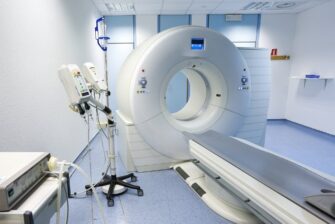
Many technologies are available to allow doctors to view organs and tissues and detect the presence of fluid or tumors. Imaging technologies help your doctor not only make a mesothelioma diagnosis but also determine a treatment plan and track your response to the treatments.
- X-Ray Imaging – Due to the prevalence of the machines and their relative low cost, X-rays are usually the first test given to many patients. X-rays do not have enough resolution to identify the cancer, but they may indicate the presence of a pleural effusion or hint at the existence of a pleural or lung malignancy.
- Computed Tomography (CT) – A computed tomography scan uses X-rays and computers to give more sophisticated and detailed pictures of the inside of the body than conventional X-rays. CT is considered the gold-standard imaging test for a diagnosis.
- Magnetic Resonance Imaging (MRI) – Magnetic resonance imaging uses a strong magnetic field and radio waves to scan the body. MRI provides much better imaging of soft-tissue contrast than does CT and is often used for staging and surgical planning.
- Positron Emission Tomography (PET) – Positron emission tomography measures functional and metabolic activity in the body. PET scans are most commonly used to identify the presence of distant metastases.
- PET-CT (Integrated Positron Emission Tomography – Computed Tomography) – PET-CT is a cutting-edge imaging technique that combines PET scans and CT scans in one machine. This allows the physician to precisely register the functional imaging of PET with the anatomical imaging of CT to provide a more complete understanding of a patient’s individual disease status.
Biopsy Procedures
Thoracentesis
Fluid is drained from the pleural area and the lung and is tested for cancer cells. This procedure is performed when a patient presents with pleural effusion, or fluid in the lungs.
Thoracoscopy
A tissue sample of a pleural or pericardial tumor can be obtained during a thoracoscopy. A thoracoscope is a telescope-like instrument connected to a video camera. It is inserted through a small incision into the chest, along with small surgical tools. The doctor can see the tumor through the thoracoscope and use special tools to take a tissue biopsy.
Laparoscopy
A laparoscopy allows the doctor to see and obtain a biopsy of a peritoneal tumor. In this procedure, a flexible tube is attached to a video camera that is inserted into the abdominal cavity via small incisions.
Bronchoscopy
With bronchoscopy, the doctor inserts a flexible lighted tube down the trachea and into the bronchi to check for masses in the airway. At the same time, the doctor may remove small samples of abnormal-appearing tissue for testing.
Mediastinoscopy
During a mediastinoscopy, the doctor inserts a lighted tube under the patient’s sternum, or chest bone, at the neck level and moves it down into the chest. The doctor can see the lymph nodes and take tissue samples to check for cancer.